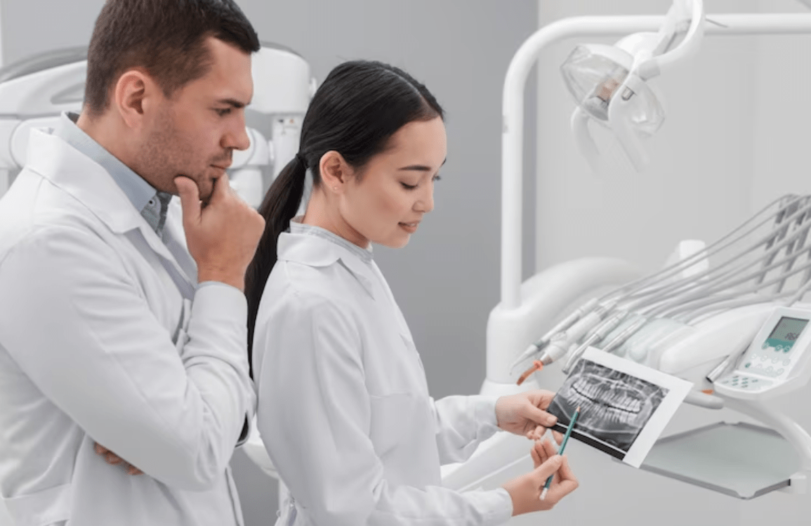
RVG is the latest modern dentistry technique for treatment processes. Modern dental imaging has gained much popularity in treating patients of late.
Accuracy in diagnosis is important in dentistry. After an accurate diagnosis, the dentist can be able to not only fix the problem but also meet the customer’s expectations. Modern dental imaging has indeed a very important role to play in dentistry. A clear image can, in fact, help in accurate diagnosis, while an unclear image can rather increase the chances of an error. Capturing a quality image is not really as simple as it sounds. Image quality is, of course, affected by a number of factors.
RVG, or Radiovisiography, happens to be the most recent technique of imaging used in dentistry. The radiation exposure is minimal as compared to the other traditional techniques used for capturing images.
Radiovisiography (RVG) happens to be the latest imaging technique in dentistry, with minimal radiation exposure to the patient as well as numerous possibilities to process the images. It also has several advantages over classic radiography.
What is an RVG sensor?
RVG means Radiovisiography and is considered a technology that has replaced traditional x-ray radiology methods. It is now extensively used in dentistry on account of its benefits.

What are the advantages of the RVG sensor?
- RVG does reduce the patients exposure to radiation by 80%.
- The sensor does help the doctor get instant results.
- There is hardly any sort of wait time for the patient’s reports.
- The sensor of the RVG is rather capable of versatile applications. It is, in fact, covered with housing and also has a shock protection layer that protects the sensor from bites as well as drops.
- The RVG is indeed capable of taking high-quality images due to its high-sensitivity scintillator, fiber optics, and high-resolution rugged CMOS detector. The digital sensor does deliver images of the highest resolution, more than 20 LP/mm.
- RVG sensors have the functionality of saving images and also changing their size or contrast for better viewing.
- RVG sensors are no doubt available in three sizes, depending on their applications, which range from 0 to 2. For pediatric examinations, size O is used, while for general purposes, size 1 is recommended. Size 2 is used for obtaining bitewing images and also.
- Teeth problems can be detected early, and the necessary steps can be taken in time to arrest the damage.
- It is very cost-efficient as it does not really involve the chemical processors.
- The soft tissue as well as fractures are easily visible due to the high quality of the image produced, which would otherwise be rather undetectable.
- With RVG, the storage of images on computers is indeed possible, and they can be shared with experts over the net and viewed on any sort of device.
- Specific areas can be enlarged in order to visualize instrument locations, especially in endodontic treatments.
- It is convenient for the radiologist to add his observations to the image.
- The radiologist can retake an image almost instantly.
- The high-quality digital images can, in fact, be easily shared with the concerned departments, thus making the entire process highly efficient.
Conclusion
Thus, the dawn of the digital era in dental radiography came in 1987 with the first digital radiography system known as Radiovisiongraphy (RVG). A physicist and also a charge-coupled device (CCD) image sensor design engineer, Paul Suni, did create the CCD image sensor technology that made the RVG digital radiography system a reality. The main factor that distinguishes digital systems from conventional ones is their response to incident radiation. A modern dental imaging system ends up operating between a completely bright and a completely dark image.

